

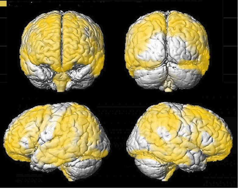
Children diagnosed with sleep apnoea but not undergoing CPAP therapy may exhibit delayed brain development. A study led by Dr Leila Kheorandish-Gozal of the University of Chicago indicated that children suffering from moderate to severe obstructive sleep apnoea could have damage to brain cells. The test subjects showed a significant reduction of grey matter brain cells involved in memory, movement, speech, emotions, self-control, decision-making, and perception when compared with children of the same age who do not have sleep apnoea. Statistics indicate that up to 5% of children are affected by sleep apnoea. This study finally linked sleep apnoea and delayed neuronal growth in a child’s developing brain. The photo below shows the brain of a child with sleep apnoea (left) and the one on the right is the brain of a child who sleeps normally. Notice the lack of grey matter on the left brain compared to the right brain. The reduction of grey matter in children with a treatable sleep disorder is quite extensive, making it imperative for parents of children who exhibit symptoms of sleep apnoea to consult with a sleep physician.
The study involved 16 children diagnosed with obstructive sleep apnoea. Each child had non-invasive magnetic resonance imaging (MRI) of his/her brain and underwent neuro-cognitive testing at the University of California Their sleep patterns were assessed at the paediatric sleep laboratory of the University of Chicago. The results of the tests were compared to the scans, MRI images and neuro-cognitive test results of 9 children of the same gender, age, weight, and ethnicity but without sleep apnoea. The results of the 16 test subjects with sleep apnoea to the MRI scans of 191 children who were in the National Institute of Health database. Multiple brain regions of the children with sleep apnoea showed a decreased volume of grey matter. These regions are the temporal lobe (hearing), prefrontal cortices (personality, planning, complex behaviour), frontal cortices (problem-solving, language, movement, memory, impulse control, judgment), and the brainstem that controls the body’s respiratory and cardiovascular functions. The researchers stated that though the images showing the changes in the grey matter are remarkable, an exact guide to link specific cognitive loss and grey matter deficit is yet to be established. However, there is a distinct indication that children with untreated obstructive sleep apnoea show neuronal damage when compared to children with no sleep disorder. The researchers stated that the direct consequences of the loss of grey matter can be difficult to measure. One of the lead researchers, Dr. David Gozal, said that the MRI scans do not show what became of the affected neurons. It cannot be determined if the affected brain cells are gone forever or just shrunk. Though the occurrence of the damage cannot be determined, the researchers can link the gravity of the sleep disorder with the range of cognitive deficits. Lead researchers from the two involved universities are planning collaborative studies to seek answers to questions raised by the initial study. According to Dr. Gozal, there is a need to test cognitive function before the beginning of sleep apnoea to quantify the effect of neuron loss. If the average IQ loss induced by sleep apnoea is 8 to 10 points, then a child with an IQ of 170 will not be greatly affected: But a child with an IQ of 100 and below will be. So far, the study indicates that untreated sleep apnoea in children could negatively impact such children.
If you have a child who is showing symptoms and signs of obstructive sleep apnoea, it is best to consult with a qualified sleep physician.
Find a clinic or call us now for a consultation at 1300 750 006.