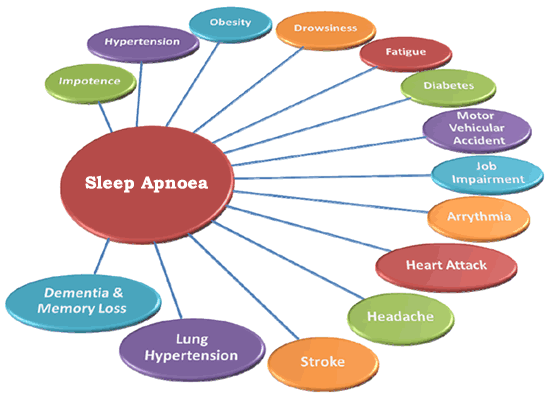
Cognitive performance is also affected. There are studies that indicate that sleep apnoea leads to slow thinking, lower intelligence quotient, poor creativeness, and brain fog.
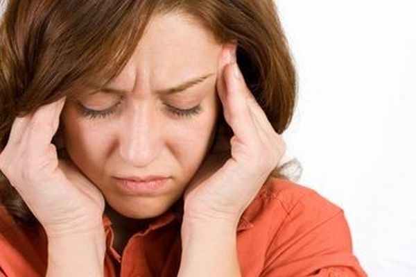
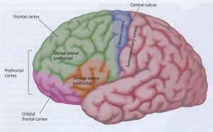
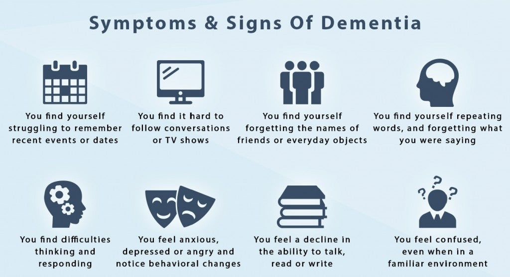
Brain fog or brain fatigue is a mental state wherein the person suffering from it exhibits poor memory recall, lack of focus and marked reduced mental acuity. This mental confusion can manifest without much warning, and the episodes can be mild to severe. If the cause of brain fog is not quickly and accurately diagnosed and treated, the episodes may reach a point wherein the person’s personal and professional life is affected.
Symptoms
Brain fog is medically termed cognitive dysfunction. A person suffering from brain fog will have difficult remembering why he came into any room. He might have to read and re-read a paragraph because he cannot remember what he read. He might be so absorbed on his daily task and not interact socially. He might feel great physically but not mentally. Remembering particular things, words, and processes could be quite a struggle, and routine tasks might require extra effort to do.
Lack of Sleep
There are various factors that may cause brain fatigue and lack of sleep is one of them. The human body needs sleep to recover from its daily tasks. The brain needs rest and sleep too in order to recuperate from the tens of thousands of processes it does during one’s waking hours. When sleep is interrupted on occasions due to sickness or late work or studies, the sleep interruption is temporary and will not likely lead to brain fog. However, if a person’s nighttime sleep is interrupted on a regularly or daily, he is more likely to experience brain fatigue each morning. There are those who shake off brain fog with a cup of espresso, but for those who are suffering from a form of sleep disorder such as sleep apnoea, brain fog cannot be easily shaken off.
Scientific studies indicate that cognitive dysfunction may be caused by chronic sleep interruptions or difficulties across all age brackets. This means narcolepsy, insomnia, circadian rhythm disorder, restless leg disorder, shift work sleep disorder, and more, are also risk factors for brain fog. Studies also indicate that brain fatigue in patients with obstructive sleep apnoea (OSA) is quite common. This could be explained by another study suggesting that diminished oxygenation of the blood is the main culprit for memory problems and physical deficiencies.
Stress is known to negatively affect the body, which could lead to sickness, including brain fog. An average person should be able to handle normal daily stress. If he starts to show brain fog, then the possibility of an underlying condition becomes real.
Neurological Disorders that may lead to brain fatigue include lupus, fibromyalgia, multiple sclerosis and chronic fatigue syndrome.
Diabetes causes glucose levels in the blood to fluctuate. Since glucose is the main energy source of the brain, brain fog may occur.
Menopause causes hormones to fluctuate thus women going through this phase may experience brain fatigue.
Deficiencies in nutrients and side effects of particular medications may also cause brain fatigue.
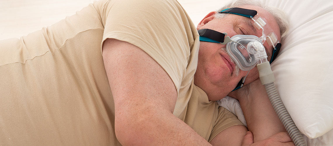
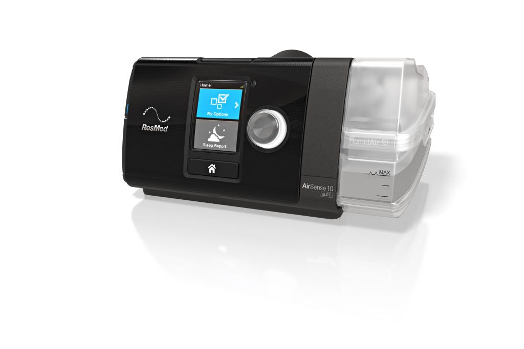
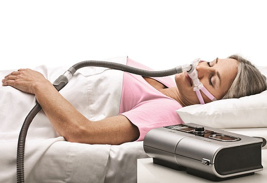
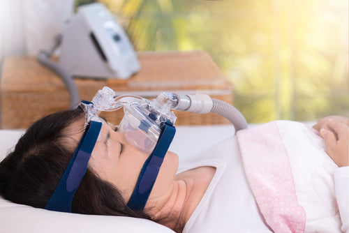
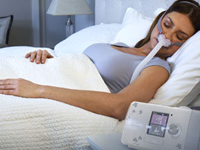
It is possible to treat brain fog. A study showed that 20 patients with severe OSA manifested brain fog. After undergoing CPAP therapy, their brain fatigue improved significantly.
If a person lacks sleep, then he should get to the root of his sleeping disorder to explore possible treatment. If the interrupted sleep is due to sleep apnoea, then consulting with a sleep doctor for correct diagnosis and treatment approach. In such case, CPAP therapy is the correct treatment.
Call us now for consultation. 1300 750 006
]]>What Causes Sleep Apnoea?
Obstructive sleep apnoea is due to the collapse of a part of the airway starting from the nose and ending up on the lungs, collapses during sleep. Breathing is repeatedly disrupted for 10 seconds or more all through the night.
The most common cause of obstructive sleep apnoea (OSA) in adults is obesity and excess weight, which is linked with the soft tissue of the throat and mouth. When the muscles of the tongue and throat are more relaxed during sleep, the soft tissue may cause blockage in the airway.
Chronic snoring causes vibration of the airway and eventually damage it, contributing to the swelling along the narrow air passage. This may lead to less responsive nerve receptors and weaker muscle tone for airway support.
Sleep position is also a contributing factor. Sleeping in a supine position may cause the tongue to fall back into the airway. Too much intake of alcohol a few hours before sleep may cause the muscles of the airway to collapse. In children, enlarged adenoids or tonsils, dental conditions, birth defects ( Pierre-Robin syndrome and Down syndrome), may cause OSA. These contributing factors are to be considered when assessing if sleep apnoea can eventually go away.
Sleep Apnoea: A Chronic Condition
Obstructive sleep apnoea is a chronic condition. Human anatomy is fixed unless an adult with OSA opts for surgical procedure. Adults with OSA may opt for a jaw advancement surgery called maxilla-mandibular advancement. This is a major surgical procedure that involves breaking the jaw bones, moving them forward and screwing them in placing titanium plates and screws. This procedure changes the facial profile, and has been proven to be at least 80% effective in resolving OSA.
Surgical procedures such as uvulopalatopharyngoplasty (UPPP) and septoplasty for repairing deviated septum are less effective in treating sleep apnoea. Hyoid advancement or hyoid suspension is a sleep surgery where the hyoid bone and its muscle attachments to the airway and tongue are pulled forward to try to increase the size of the airway and improve airway stability behind and below the base of the tongue.
Children with OSA may hope for the condition to go away if the cause is due to enlarged tonsils and adenoids as tonsillectomy ad adenoidectomy are the answers. For sleep apnoea due to dental conditions, orthodontic procedures will prove very helpful.
Changing Sleep Apnoea Risk Factors
Not everyone with OSA, for various reasons, will opt for surgical procedures. However, there are other ways to reduce the presence and/or severity of sleep apnoea. Overweight people with OSA can lessen the severity of their condition by losing weight. Fat tissue that lines the airway contributes to narrowing the airway and its collapse. Losing weight, and sleeping on one’s side instead of in supine position, will definitely have a positive effect. It is best to sleep on one’s side, with the head propped from 30 degrees to 45 degrees.
Tongue exercises such as myofunctional therapy and playing the didgeridoo for toning the airway muscle will help improve sleep apnoea. However, aging causes lost of muscle tone and without treatment, will worsen OSA. In cases where there are no other feasible options for treating OSA, continuous positive airway pressure (CPAP) therapy is still the best option.
Call us today at CPAP Victoria.
]]>
Sleep apnoea could be considered as a potentially critical sleep disease that causes a patient to repeatedly stop breathing for short periods during sleep. Sleep apnoea is one of the most common triggers for the development of pulmonary hypertension.
Sleep Apnoea and Pulmonary Hypertension
Sleep apnoea comorbidities include diabetes, hypertension, ischemic heart disease, vascular disease, stroke, arrythmias and congestive heart failure. Thus, sleep apnoea is also a common denominator for the development not of only pulmonary hypertension but also cardiovascular disease. Research indicates that hypercapnia (elevated carbon dioxide in the body) repetitive nighttime arterial oxygen desaturation (low blood oxygen concentration) are the two main factors that contribute to the increase in pulmonary artery pressure.
An adult needs at least a full 8-hour of uninterrupted sleep for his body to repair and mend damage to the cellular level. Even the human growth hormone (HGH) is only release during deep sleep, which is achievable only during long periods of consolidated sleep.
Constant lack of sleep, or poor sleep greatly diminishes the body’s ability to recharge and repair. Obesity and aging are common risk factors for developing PH. Aging cannot be controlled while obesity can be effectively controlled and managed. If obstructive sleep apnoea (OSA) or any related sleep-disordered breathing condition, remains undiagnosed and untreated, the development and eventual progression of PH is a certainty.
Treatment of OSA and PH
According to researchers from the American College of Chest Physicians, between 17 and 53 percent of people with sleep apnea also develop pulmonary hypertension. The repeated loss of oxygen in the bloodstream due to OSA and other variants of sleep apnoea increases the pressure of the pulmonary artery.
Any 10-second, or longer, cessation in breathing during sleep is considered an apnoea. The frequency and length of apneas determine the amount of loss of oxygen in the blood. Any significant loss of blood oxygen will increase the carbon dioxide level in the blood. The decrease and increase of oxygen and carbon dioxide in the blood will stress the cardiopulmonary system, which could lead to other complications such as pulmonary hypertension. The cessation of breathing during sleep also causes negative changes to internal lung pressure, which could affect the heart.
A person diagnosed with PH can opt to have a sleep study to determine if his PH is linked to undiagnosed or untreated obstructive sleep apnoea.
Treatment for OSA- related PH is through the use of continuous positive airway pressure (CPAP). Studies have indicated that CPAP therapy can efficiently reduce pressure in the pulmonary artery.
Obesity is a controllable risk factor of both OSA and PH. An obese person with both OSA and PH must reduce his weight.
If you or any of your family has OSA or sleep apnoea-related PH, call us now.
1300 750 006
]]>Published in the Journal of the American Heart Association, the study explored the link between sleep apnoea and heart failure. Sleep apnoea is a condition wherein a person stops breathing over 5 times per hour during sleep.
Previous scientific research and studies have indicated that people with untreated obstructive sleep apnoea (OSA) are at risk of developing comorbidities such as type 2 diabetes, hypertension, stroke, arrhythmias, and heart attack. This recent study focused on the connection between heart failure and sleep apnoea. Heart failure is not the same as a heart attack. Heart failure is when the heart is not pumping well. When this occurs, the body receives less oxygen.
Among the 4.9 million Danish adults the researchers looked over 12 years (2000 -2012), 40,4085 developed sleep apnoea during the study. The researchers found that patients with sleep apnoea who are not undergoing CPAP treatment have a slightly higher risk of developing heart failure, regardless of their age. The study also indicated that those over 60 are 38% more likely to suffer heart failure than those who use CPAP.
Dr Anders Holt, the lead author of the study and a researcher with the Department of Cardiology at Copenhagen University, said that there is no denying that people with sleep apnoea are more prone to heart failure than the general population. Holt expressed that doctors caring for patients with sleep apnoea should pay close attention to observing and treating not only sleep apnoea but other cardiovascular risk factors.
The result of this study was reinforced by Dr. Donna Arnett when she said that the use of a CPAP machine is a positive step against the development of heart failure. Dr. Arnett is currently the dean of the College of Public Health at the University of Kentucky. Though she was not part of the study she believes that there is a great need to educate physicians about the significance of sleep apnoea. She said that the sleep disorder is under-recognized and that patients should be encouraged to get screened.
Dr. Hold admits that the current research was limited due to a lack of information about cardiovascular risk factors such as body mass index, smoking and drinking status. He hopes that further studies will be randomised clinical trials. However, the study supported the efficacy of CPAP therapy in decreasing the threat of developing heart failure for sleep apnoea patients. It underscored the benefits of CPAP therapy for patients with known heart failure.
The study highlights the importance of helping patients with sleep apnoea to adjust to sleeping while undergoing CPAP therapy.
Find a clinic or call us now for a consultation 1300 750 006.
]]>According to NICHD’s Pregnancy and Perinatology Branch’s Uma Reddy, M.D., the study opened the door for very affordable means to screen and test large numbers of women who are at a higher risk of developing sleep apnoea during pregnancy. Dr. Reddy said that the screening method will facilitate a quick way to identify who required further tests. She added that future studies may lead them to find ways to improve the outcomes of pregnancy.
An earlier study of first-time pregnant women diagnosed with sleep apnoea indicated that the risk of developing gestational diabetes and hypertensive disorders is higher for them. There are NIH-supported studies of potential treatment for pregnancy-related sleep disorders but as of now, there are no treatment recommendations or medical guidelines for sleep apnoea during pregnancy. The Maternal Foetal Medicine Units Method has funding from the NICHD and is planning a larger study about pregnancy-related sleep apnoea.
The subjects of this sleep study answered questionnaires regarding their daytime sleepiness, snoring, and sleep habits during their early pregnancy stage of 6 to 15 weeks, and mid-pregnancy stage of 22 to 29 weeks. They also underwent at-home sleep apnoea testing.
The result of the study indicated that 3.6% of 3, 264 women in early pregnancy and 8.3% of 2, 512 women in mid-pregnancy have developed sleep apnoea. The study pointed out that obesity based on body mass index (BMI), older age and frequent snoring are risk factors.
The authors of the study have developed a method of calculating the probability of developing sleep apnoea in early and mid-pregnancy based on a woman’s BMI, maternal age, and snoring frequency. This method or tool is being made available to obstetricians and other related providers to be able to identify pregnant women who are at risk, and in need of further testing.
The gold standard for treating sleep apnoea is continuous positive airway pressure or CPAP therapy. The treatment therapy involves wearing a mask that fits over the mouth and nose, or nose only. A tube is attached to the mask and pressurised air is pumped through it to keep the airway from collapsing. Dr. Uma Reddy added that it is not known yet if CPAP therapy during pregnancy could prevent diabetes, hypertension, and other co-morbidities of sleep apnoea. The best recommendation for pregnant women with sleep apnoea is to see a qualified sleep doctor to assess her condition and treatment in consultation with her obstetrician.
Find a clinic or call us now for a consultation at 1300 750 006.
]]>
PSG is a non-invasive procedure. The sensors used may cause minor skin irritations to some, but the discomfort is insignificant. The comfort and safety of patients during the testing are ensured as trained and qualified staff are on duty during the diagnostic test.
Physiological processes that are measured during a PSG diagnostic test include: electrocardiogram (ECG), electroencephalogram (EEG), electrooculogram (EOG), electromyogram (EMG), nasal and oral airflow, leg movements, snoring and other sounds, and position of the body.
There are several types of sleep studies available.
Diagnostics Sleep Studies
Diagnostic Sleep Studies include overnight and daytime diagnostics. An overnight diagnostic PSG sleep study is the most common. It involves non-invasive sensors attached to key points to diagnose obstructive sleep apnoea (OSA), snoring, sleep-state misperception, limb movement, and other sleep disorders. This test also helps in qualifying narcolepsy, insomnia, restless leg syndrome and idiopathic hypersomnolence. For patients whose work calls for them to work during the day, a daytime diagnostic sleep study is done.
MSLT or Multiple Sleep Latency Test
This is a daytime sleep study favoured for diagnosing hypersomnolence and narcolepsy. An MSLY is usually done following an overnight PSG. The diagnostics entail from 4 to 5 periods of napping at intervals of 2 hours during the day. This test measures and records eye movements and brain waves to diagnose sleep stages and sleep times.
Electroencephalograms or EEG
This test will determine and measure convulsions, seizures, and other abnormal movements during sleep. The test is recorded and analysis of the results by a qualified physician is done after the EEG.
MWT or Maintenance of Wakefulness Test
This test is for assessing a patient’s ability to stay awake during the day. It is usually done after a patient has undergone or is undergoing treatment for his sleep disorder. Done after overnight PSG, MWT calls for four 40-minute test episodes on intervals of 2 hours during the day. Eye movements and brain waves are measured to determine the patient’s sleep state and wakefulness. This test is most helpful for determining if a patient is safe to drive.
CPAP, BiPAP, APAP Titration Studies
A CPAP titration test is for determining the CPAP pressure of patients prescribed to undergo CPAP therapy. The correct CPAP pressure is needed to control obstructive respiratory episodes and snoring. Sensors much like the ones used in PSG are used for a titration test. BiPAP and APAP titration studies are like CPAP titration studies, as they also determine the correct BiPAP or APAP pressure for effective therapy.
Find a clinic or call us now for a consultation at 1300 750 006.
]]>The study indicated that changes in the brain during sleep apnoea episodes are directly linked to changes in the structure of the brain seen in elderly people with signs of early dementia. Scientists at the University of Sydney’s Brain and Mind Center said that new findings indicate that oxygen deprivation during sleep could be tied to alterations in the brain’s temporal lobes and compromised ability to absorb new information.
The lead researcher, Sharon Naismith, said that results of the study pointed out that oxygen desaturation during sleep is a probable contributing factor in changes in the brain and memory of elderly people with sleep apnoea. Statistics indicate that about 75% of Australiana over the age of 65 are affected by obstructive sleep apnoea. Treatment of the sleep disorder could help prevent the occurrence of dementia in the said age bracket. One of the recommendations of the study is that elderly people should be screened for obstructive sleep apnoea.
OSA and Dementia
There are over 50 million people across the globe who gave been diagnosed with dementia. As a syndrome, dementia causes the afflicted to suffer a decline in their ability to remember, think, converse and do everyday things to live independently.
OSA has been tied to heart disease, stroke, high blood pressure, diabetes, cancer and now, dementia. Up to 70% of dementia cases are due to the onset of Alzheimer’s. A 2017 study indicated that the build of toxic protein int the brain relating to Alzheimer’s is directly linked to raised amyloid beta due to obstructive sleep apnoea.
OSA and Alterations to Brain Structures
The study spearheaded by Professor Sharon Naismith involved 83 individuals with ages ranging from 51 to 88. All of the subjects have consulted with their personal doctors regarding mood and memory problems. Not one of them has been diagnosed with OSA.
All of the subjects went through depression symptoms screening, tests for memory ability, MRI and OSA assessment that included polysomnography. The polysomnography test allowed the researchers to see changes in blood oxygen, brain activity, heart rate, breathing, while the MRI scans allowed the researchers to measure the various parts of the subjects’ brain.
The tests established that low levels of blood oxygen during sleep is directly linked to decreased thickness of the brain’s left and right temporal lobes, which are the brain structures that are fundamental for memory and are identified to change in dementia. The same tests showed that the changes were closely connected with diminished verbal encoding for retaining new information.
OSA and CPAP
Dementia cannot be treated, but depression, obesity, high blood pressure, smoking and even OSA are modifiable factors.
Obstructive sleep apnoea can be treated with continuous positive airway pressure (CPAP). The research team has embarked further studies to investigate the role of CPAP therapy in improving brain connections and put off cognitive decline in people with mild cognitive impairment which may or may not lead to dementia.
If you or someone you care about has sleep apnoea, call us. We can help! 1300 750 006
]]>
A study about comorbid insomnia and obstructive sleep apnoea indicated that 39% up to 58% of OSA patients are also suffering from insomnia. The study showed that 43% of elderly people diagnosed with chronic insomnia have undiagnosed OSA.
The question now is does a person have sleep apnoea because of insomnia or does a person have insomnia because of sleep apnoea?
Study #1
To establish a link between insomnia and sleep apnoea, Dr. Barry Krakow and his team conducted a study with the objective of finding an answer to why an insomniac wakes up at night. From the group of potential subjects of the study, all those who appear to have sleep apnoea were eliminated. Twenty subjects who gave no indication of suffering from sleep apnoea passed the screening and became the subjects of the study.
The results of the study showed that out of the 20 subject/patients, 18 are suffering from sleep disordered breathing, affecting them to wake up during sleep. Of the 20 subjects/patients, 11 have moderate obstructive sleep apnoea.
The conclusion of the study indicated that sleep apnoea and insomnia are linked. The recommendation is for insomniac who wake during the night should get tested for sleep apnoea.
Study #2
After extensive research about underlying insomnia in patients with sleep apnoea, the director of Behavioral Sleep Medicine in Columbia, Maryland, Dr. Emerson Wickwire, said that there is evidence that insomniacs have a high occurrence of sleep disordered breathing and that sleep apnoea patients have an increased risk of developing chronic insomnia.
Dr. Wickwire also said that people with insomnia have narrow upper air passageways, more so behind the tongue. These people may either have undiagnosed sleep apnoea or may be suffering from a condition known as upper airway resistance syndrome or UARS, a condition where the breathing pause is not long enough to indicate apnoea. However, the patient does go through numerous breathing pauses, which creates stress response, causing the insomniac’s to respond negatively and worry incessantly about everything, which further aggravates the insomnia.
Conclusion
Based on the two studies done by experts in the field of sleep disorder, it is best for an insomniac who wakes up during the night to consult with a qualified sleep doctor in order to rule out obstructive sleep apnoea. An OSA patient who has insomnia should also seek medical help to address the underlying cause of insomnia.
Continuous positive airway pressure (CPAP) therapy remains to ne the most effective way to treat obstructive sleep apnoea.
Call us! We can help you address your sleep disorder and/or insomnia.
]]>In the last 25 years, continuous positive airway pressure (CPAP) therapy has been the gold standard for treating sleep apnoea. However, not everyone prescribed with CPAP therapy can tolerate the treatment.
Alternative Sleep Apnoea Treatments
In past blog articles, CPAP Victoria has presented alternative treatment for CPAP therapy. Alternative treatments for mild cases of OSA include positional therapy and lifestyle changes such as losing weight and regulating alcohol consumption.
Articles promoting change of diet, doing tongue, mouth and throat exercises and playing the didgeridoo to strengthen airway muscles were also featured in blog articles. The use of the mandibular advancement device (MAD), an oral appliance, may be recommended by a sleep doctor through a qualified dentist. Surgical procedures such as tracheostomy, tonsillectomy, adenoidectomy, bariatric for weight loss, and removal of excess throat tissue in the airway are not routinely prescribed, and are therefore considered last resort treatments if all other treatment options prove to be ineffective.
What other natural ways and non-invasive procedures can help “treat” sleep apnea?
Singing actually strengthens airway muscles, thus, theoretically, prevent those muscles from collapsing during sleep. A study shows that singing for 20 minutes each day for at least three months actually reduced snoring. The vocalization should entail singing vowel sounds such as ah and yah with wide open mouth, opening the back of the throat. The tongue should be positioned so as not to block the air passing back and forth over the throat. Singing is one harmless, fun, inexpensive, and enjoyable way to treat sleep apnoea.
Yoga has been known to help people deal with various types of health concerns for over a hundred years. Yoga may not actually treat sleep apnoea but doing yoga exercises could help alleviated sleep apnoea symptoms. Sleep apnoea is synonymous with improper breathing and subsequent depletion of oxygen levels in the body which would lead to fatigue, depression and other comorbidities. By practicing yoga, you strengthen the diaphragm and consequently increases the body’s oxygen level. Yoga is a calming exercise that could help you have a deeper and undisturbed nighttime sleep.
Acupuncture has been used to treat hundreds of ailments since the time of Ancient China civilization. The use of ultra-fine needles to puncture the natural meridian lines of the body has been documented to “cure” illnesses, and alleviate pains and aches. Acupuncture needles are painless. There are specific puncture areas for specific ailments, stimulating hormones to activate and heal the body and in the process contribute to easing the symptoms of sleep apnoea. There are actual scientific studies that show the viability of acupuncture in the treatment of obstructive sleep apnea. The finding is that acupuncture stimulates the throat’s upper airway, keeping it open during sleep for better intake of oxygen and easier breathing.
Tried and True Therapies
The researchers of the American College of Physicians Clinical Guidelines Committee have reviewed studies pertaining to the effects of the different approaches in the treatment of obstructive sleep apnoea. According to the team of scientists, weight loss and CPAP therapy are still the best options in the treatment of obstructive sleep apnoea (Epstein, 2018).
Call us NOW at 1300 750 006 we can help you!
]]>How many are sleep deprived?
Studies indicate that in developing countries, about 150 million people are sleep deprived. In the United States alone, 1/3 of the population is not getting enough sleep. In Japan, the average sleeping hour is only 5 hours and 59 minutes. According to research done by the Sleep Health Foundation 33%-45% of Australiana have poor sleep patterns that lead to irritability and fatigue that could lead to unsafe behavior, poor mental health and low productivity.
Sleep deprivation could be caused by various factors; but one of the most common causes of poor sleep quality is sleep apnoea. People with undiagnosed and untreated sleep apnoea may develop comorbidities such as heart disease, diabetes, hypertension, asthma, GERD, depression and more.
Treatment Options
Sleep apnoea such as obstructive sleep apnoea or OSA is highly manageable. The gold standard treatment for OSA patients is the use of a continuous positive airway pressure or CPAP machine. For sleep apnoea patients who cannot tolerate CPAP machine, other treatment options such as dental appliance, upper airway surgery to remove excess airway tissue, positional therapy, and even lifestyle changes such as losing weight and quitting drinking and smoking. But for OSA patients who have tried and failed using a CPAP machine, there is a new alternative – the hypoglossal nerve simulation method.
What is a hypoglossal nerve stimulation?
The hypoglossal nerve is the cranial nerve that controls tongue movement. It has a motor function and is a breathing sensor as well. A hypoglossal nerve stimulator (neuromodulation device) is like a pacemaker as it is implanted in the chest of the patient. The hypoglossal nerve stimulator is FDA-approved and as of this writing there are medical companies that have started to manufacture the device and at least one brand is now available in the market. Also called a neuromodulation device, the innovation received the top 2 award for the Top 10 Medical Innovations for 2018.
https://youtu.be/cu1AeHUZIuY
The hypoglossal nerve stimulator has leads that are connected to the hypoglossal nerve. The initial lead is positioned in the chest to detect when the patient inhales. The secondary lead is placed in the tongue, nudging it forward as the patient breathes. The mild stimulation of the tongue causes it to move and as a result open the airway, clearing any obstruction that could cause apnoea.
Clinical Trial
As of this writing, there is about 2,500 patients with sleep apnoea have opted for a hypoglossal nerve stimulator implant. These patients did try CPAP therapy but all have various reasons for not adhering to the therapy. OSA patients and their loved ones are truly negatively impacted with untreated sleep apnoea. Opting for the hypoglossal nerve stimulator implant seems to be their last resort. Clinical trials indicate an adherence rate of 6 to 7 hours of sleep for the tested stimulator brand. Requirements to qualify for the implant are:
If you or anyone you care for is suffering from obstructive sleep apnoea, contact us now! We can help you each step of the way to a successful CPAP therapy.
1 300 750 600
]]>People with obstructive sleep apnoea experience at least 10 seconds of restricted breathing during sleep due to blockage in their airway. The condition can be mild or severe, depending on the number of times a person stops breathing each hour.
What is Atrial Fibrillation?
Atrial Fibrillation or AFib is a condition that is characterized by an abnormal heart rhythm experienced by a person due to disordered electrical signal received by the heart. During AFib the atria or upper chambers of the heart quiver instead of contract. The blood pools in the atria which could form into clots that break free, enter the bloodstream and cause a stroke.
Clinical studies have shown that people with AFib are five times more at risk of having a stroke. People with diabetes, hypertension, heart disease, and those over 60 are more at risk of developing AFib. Until recently, the link between sleep apnoea and AFib has not been fully understood. It is only now that health care providers are aware that AFib and ISA are two overlapping conditions – that addressing both conditions is the best treatment approach.
OSA and AFib Relationship
Statistics indicate that there are more than 100 million people around the world suffering from sleep apnoea. Research indicates that people with sleep apnoea are 4 times more likely to develop atrial fibrillation. If sleep apnoea remains untreated, then the onset of comorbidities such as diabetes and hypertension are likely to develop, inclining a person to develop AFib. So, these two conditions are closely linked and connected.
Further research indicated that sleep apnoea triggers arrhythmias while one is sleeping. A person with sleep apnoea lacks oxygen for several seconds numerous times each hour. Lack of oxygen causes the heart to undergo chemical changes and mechanical stresses, which may consequently lead to atrial fibrillation. Untreated sleep apnoea may inhibit AFib treatment because the condition lessens the efficacy of some AFib treatments. For example, patients with both OSA and AFib may not respond well to heart medications compared to patients with AFib only. Thus, a patient with AFib and OSA will respond more with his AFib medications if his sleep apnoea is under control.
Treating OSA and AFib
A patient with AFib and OSA should be treated accordingly. The usual approach for treating AFib is by a minimally invasive technique called catheter ablation. The procedure involves threading a catheter through the blood vessels and to the heart’s left atrium. A cryo energy or radio frequency cauterizes the heart muscle that short circuits the hearts electrical system, thus stopping any atrial fibrillation. This procedure will be highly effective in combination with CPAP therapy for patients with both OSA and AFib.
CALL US NOW FOR MORE INFORMATION ABOUT SLEEP APNOEA!
1 300 750 006
]]>Obstructive sleep apnoea is characterized by intermittent breathing during sleep. It is a sleep disorder that is linked to variety of problems not only in people with diabetes but to non-diabetics as well. OSA is associated to poor work performance brought about by disturbed sleep. OSA comorbidities include hypertension, arrythmias, stroke and even heart failure. A diabetic person with sleep apnoea may even develop insensitivity to insulin. With millions of people across the globe suffering from sleep disorder and diabetes, the risk for cardiovascular death has greatly increased.
Interaction between Sleep Apnoea and Diabetes
There are several mechanisms that link sleep apnoea and diabetes.
Inflammatory Reaction. Systemic inflammation, inflammation of the upper airways and inflammation of the lining of the blood vessels are associated with sleep apnoea. Obesity, high levels of triglycerides and cholesterol in the blood, cardiovascular disease and atherosclerosis are also linked to systemic inflammation.
Stress Reaction. The body is triggered to a fight or flight mode if it repeatedly deprived of oxygen due to sleep interruption caused by sleep apnoea. The stressor increases blood pressure and heart rate which could lead to cardiovascular disease, insulin resistance and chronic high blood pressure.
Lack of Oxygen. Repeated oxygen deprivation may prod the body to produce proteins associated with insulin resistance and glucose intolerance.
Cortisol Levels. Consistent sleep interruption may increase the cortisol level, leading to raised insulin secretion and blood glucose levels.
Link to Syndrome X
Syndrome X or metabolic syndrome is linked to sleep apnoea and type 2 diabetes. The syndrome is associated to five medical conditions that increases the risk of diabetes and atherosclerosis. The five medical conditions are high blood fasting glucose level, high blood pressure, abdominal fat, low HDL or food cholesterol, and high triglycerides.
Raised fasting glucose. Obstructive sleep apnea is linked with insulin resistance and glucose intolerance regardless if a person is obese or not. The condition has also been linked to insufficient sleep, interrupted sleep, and difficulty in sleeping and maintain sleep – all of which are associated with sleep apnoea and snoring
High Fat. Studies indicate that excess body fat in the abdominal area is a good indicator of sleep apnoea. Research shows that about two-thirds of people who snore or have sleep apnoea are obese. As the body-mass index (BMI) increases, the severity of OSA increases.
High blood pressure. The risk of high blood pressure also increases with increasing severity of sleep apnea, and it has additionally been linked with insulin resistance.
Low HDL and Triglycerides. People with type 2 diabetes commonly have reduced level of HDL or good cholesterol and elevated level of triglycerides, independent of BMI but relating to even a mild form of sleep apnoea.
OSA is independently linked to each of the five syndrome x conditions. In relation, the syndromes could be aggravated by untreated diabetes and sleep apnoea.
Treatment
Diabetic, OSA patients treated with CPAP therapy improved in management of their diabetes. This is an indication that sleep apnoea does have a part in the changes brought about by diabetes. A 1994 study of a group of men and women with OSA and type 2 diabetes received CPAP treatment for four months. After the prescribed length of therapy, the group’s sensitivity to insulin significantly improved.
A 2004 study gauged the effects of CPAP therapy on insulin sensitivity in a group of diabetic people with sleep apnoea. After three months of therapy, their sensitivity to insulin improved.
A 2005 study of 25 people with sleep apnoea and diabetes received a 4-hour CPAP therapy for four months. The study indicated that the group had improved blood sugar levels after meals, with lower HbA1c or glycosylated hemoglobin.
These studies reinforce the supposition that CPAP therapy is an efficient and effective component in the treatment of which the end goal is to improve the control of a patient’s blood sugar level, and to normalize his sleeping pattern.
Call us now for more information on OSA and CPAP therapy.
]]>Dangers of Bruxism
Teeth grinding is damaging to oral and dental health. Undiagnosed and untreated teeth grinding will ultimately lead to gum disease and tooth loss. Occasional teeth grinding may not be harmful, but when the condition is severe, dental damage is inevitable as well as facial pain and disturbed sleep and daytime sleepiness due to sleep fragmentation or awakenings from sleep. The condition is also associates with sleep apnoea, snoring and other night-time movement disorders.
Causes of Bruxism
The causes of bruxism are unspecified but there are studies that link the condition to alcohol consumption, cigarette smoking, caffeine and fatigue. If any one of these is the cause of a person’s bruxism, then a change in his lifestyle may well cure his condition. However, bruxism is also linked to snoring, sleep apnoea or jaw misalignment, professional help is required. Evidence indicates that a person with bruxism and sleep apnoea was able to alleviate his teeth grinding when his sleep apnoea was addressed.
Risk Factors
Bruxism has other risk factors that include one’s personality type, medications, family members with bruxism, age, and other mental and medical health disorders such as dementia, Parkinson’s disease, epilepsy, gastroesophageal reflux disorder (GERD), attention deficit hyperactivity disorder (ADHD), night terrors and other sleep-related disorders.
Signs and Symptoms
Some of the most common signs and symptoms of bruxism are:
Diagnosis and Treatment
A clinical diagnosis is done based on the history and symptoms of a patient. A certified dentist or oral care provider usually provides evidence-based diagnosis founded on the damage to the patient’s teeth. Polysomnography is not initially required but a sleep study is best for assessing if the bruxism is in anyway linked to sleep apnoea or any other movement or sleep disorder such as periodic limb movement disorder or restless leg syndrome. A sleep study will also determine the sleep disruption experienced by the patient during sleep. The same study will either confirm or refute the association between the patient’s nocturnal teeth grinding and clenching, and epilepsy or other seizure-related activity during sleep.
Treatment for bruxism depends on the underlying cause of the condition. If bruxism is due to too much caffeine, alcohol, smoking and late nights, then a change of lifestyle or behavioral modification may well be the solution. If the condition is brought about by sleep apnoea, then it is best to consult with a certified sleep doctor for CPAP therapy. In severe cases of stress, anxiety or movement disorder, anti-depressant or muscle relaxant may be prescribed.
Mouth guard to be worn during sleep is usually prescribed by a dentist to protect the teeth from further damage. Practicing good sleeping hygiene and habits is also highly advised.
Below is a simple infographics presentation to better explain bruxism.
Teeth Grinding Guide created by Schererville & Chesterton Family Dentistry.
Call us at 1300 750 006 for enquiries about sleep apnoea and CPAP therapy.
]]>Sleep Medications
Taking sleep medications may worsen the condition by inhibiting the airway. A study done by the US Centers for Disease Control and Prevention indicated that close to nine million people in the United States alone take prescription sleeping pills in the past month, thereby increasing the potential dangers of sleep apnoea. Why?
Episodes of sleep apnoea end when the sleeping person is awakened due to lack of oxygen, loud snoring, or any other respiratory distress signal. If a person with OSA takes sleeping pills, it will be harder for him to be jarred awake out of a deep sleep cycle, making the OSA episode continue for a longer period.
Excessive Weight
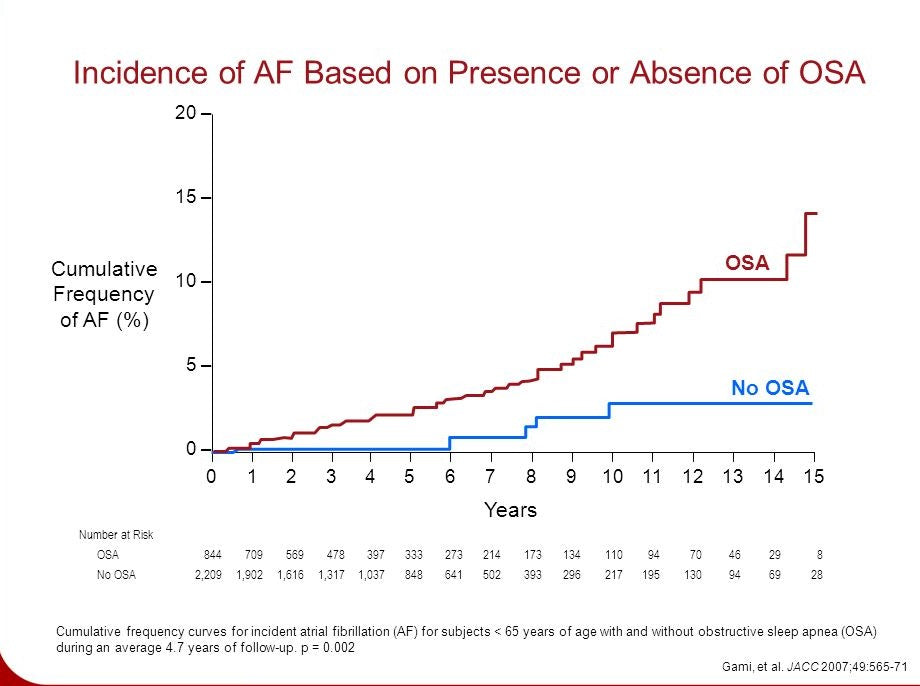
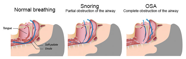
One of the most common causes of sleep apnoea is excessive weight gain as attested by a study done by the National Center for Biotechnology Information. This study qualified that more than 50% of OSA patients are overweight and that their excess weight tends to accumulate around the neck. On the other hand, OSA itself has shown to cause weight gain too. This is due to a compromised sleep cycle that wrecks the normal rhythm of the hormones that regulate hunger.
Genetics
Some people may be predisposed to sleep apnoea due to physical traits they inherited. Some of the physical attributes than can influence one’s chances of developing sleep apnoea includes shape of skull, head, oral cavities, neck circumference, and naturally narrowed throat.
Studies indicate that those with a family history of OSA are more likely to develop sleep apnoea. Birth defects such as Down syndrome and Pierre Robin sequence are also linked to sleep apnoea.
Men are twice likely to develop sleep apnoea. Women are also at risk if they are overweight. Both male and female increase their risk of developing sleep apnoea as they grow older.
Alcohol
Although drinking alcohol may help one get to sleep, the substance can worsen obstructive sleep apnoea. Why? For one, alcoholic beverages loosen the back muscles of the throat which obstructs the airway and worsen sleep apnoea symptoms. Two, too much alcohol consumption can lead to weight gain, which is another risk factor of sleep apnoea. Three, alcohol reduces rapid eye movement or REM during sleep, thus waking up a person several times during sleep.
Smoking
Studies show that smokers are three times more likely to develop sleep apnoea than non-smokers. This is due to the increased amount of fluid retention and inflammation in the upper airway caused by smoking. Quitting smoking will diminish the risk.
Sleep Position
Supine sleepers are more at risk of developing sleep apnoea, but the condition could develop for prone and side sleepers as well. Supine or back sleepers have the weight of their neck and surrounding tissues and muscles forcing the airway. This causes obstruction and restrictions on the airway during sleep, which may lead to sleep apnoea. It will be more beneficial for most people to sleep on their side.
Call us for consultation regarding obstructive sleep apnoea.
]]>Hypopnoea and Sleep Apnoea
Hypopnoea and sleep apnoea belong to the same family of sleep disorder. Where hypopnea is indicated by the partial obstruction of air during sleep or when awake, apnoea is indicated by the complete obstruction or blockage of air during sleep. Most often, these two occur simultaneously.
Doctors observed that patients with sleep apnoea sometimes experience only partial obstruction of air during sleep, instead of experiencing complete periodic obstruction. Sometimes, a combination of partial (hypopnea) and complete (apnoea) is manifested by patients. As a result of this discovery, sleep doctors concluded that a person may have hypopnea and sleep apnoea, and that a person diagnosed with hypopnea may eventually develop sleep apnoea.
What causes hypopnoea?
The causes of sleep apnoea and hypopnoea are very similar. Obstructive sleep hypopnea/apnoea is due to the relaxation of the muscles of the throat while one sleeps. Central sleep hypopnea/apnoea is due to the brain’s failure to signal the muscles to allow one to breathe.
As with sleep apnoea, there are risk factors for hypopnea. In the case of obstructive sleep hypopnea risk factors include: gender (more common in men), age, genetics, obesity, size of one’s neck, alcohol consumption, smoking, congested nasal passages, taking of sleep medication or sedatives. Other factors that could increase the risk of developing hypopnea include: heart disease, history of stroke, hypothyroidism, enlarges tonsils or adenoids in children, and other narcotics.
Are there treatment options?
As with sleep apnoea, there are treatment options for sleep hypopnea, and they depend on the severity and cause of the ailment. As with the causes of hypopnea, treatment options are the same as those of sleep apnoea.
Where hypopnoea is mild, changes on one’s lifestyle may actually decrease and eventually eliminate the condition. Lifestyle changes may include: eating a healthy diet, losing weight, smoking cessation, decreasing or completely stopping alcohol consumption, changing sleep positions, and avoidance of sedatives and other narcotics.
OSAHS or obstructive sleep apnoea-hypopnoea syndrome is a graver form of sleep hypopnoea or sleep apnoea. This condition is chronic and requires continuous management, and lifelong treatment.
Is hypopnoea preventable?
In cases of mild hypopnoea, symptoms can be eliminated by minor medical treatments and lifestyle changes. In the case of severe and chronic hypopnoea, management and treatment may take more time, and in some cases, a lifetime. However, all hope is not lost! As soon as symptoms manifest, it is best to consult with a credible physician. Early diagnosis and management will help reduce the severity of hypopoea, and the length of treatment.
Hypopnoea may not be preventable in some cases but taking steps to reduce the risks for developing hypopnoea is highly recommended.
Call us now for consultation and more information.
]]>
What is a Didgeridoo?
Didgeridoo is a 4 to 5 feet long wood wind instrument indigenous to Australia. Its origin dates back some 4,000 years. It is unlike other woodwind instruments as it is played not by using the lungs but with the movement of the tongue, lips and vocal cords. It is more of a rhythm instrument as the “beat” of the song and not its melody is played. The instrument is intuitive to learn as there is no need to know how to read notes or take special music lessons to play the didgeridoo.
Didgeridoo linked to Sleep Apnoea Therapy
It is true that each alternative treatment targets cause-specific snoring and sleep apnoea. It is also true that one of the most common causes of why an OSA patient has episodes of breathing pauses during sleep is due to his collapsed airway. If the pharyngeal muscles are exercised, will these muscles be strong and stiff enough so as not to collapse?
There are pockets of clinical studies and researches that demonstrate the efficacy of “playing” a didgeridoo to improve the quality of sleep and breathing during sleep of a person with mild sleep apnoea. The circular breathing used in playing the instrument is an efficient therapy for improving or treating snoring and mild sleep apnoea.
Sleep Apnea/ Didgeridoo Clinical Study
A 2005 study regarding didgeridoo playing as an alternative treatment for sleep apnoea and snoring was published on the British Medical Journal. The study involved 25 patients who snore and have moderate obstructive sleep apnoea. It was prodded by reports of didgeridoo players experiencing reduced snoring and daytime sleepiness.
These patients were assigned into two groups at random. One group was designated as the intervention group that was given didgeridoo lessons. For 4 months this group attended lessons and were made to practice at home. The control group was on the waiting list for lessons.
Daytime sleepiness and apnoea scores significantly improved in the intervention group, as compared to the control group. When interviewed, partners of the didgeridoo group said that their partners exhibited less sleep disturbance. Overall quality of sleep was not significantly different from the control group. However analysis of other sleep related measures indicated moderate to big effect of didgeridoo playing.
It was concluded that regular training of the upper airways by playing the didgeridoo will definitely reduce snoring and daytime sleepiness in patients with moderate obstructive sleep apnoea. The partners of the patients also exhibited improvement in their sleep quality.
Didgeridoo playing will not be effective for:
Conclusion
Didgeridoo sleep apnoea therapy may or may not work for everyone. In case it does work it is best not to assume that you OSA is treated to the point that you do away with your CPAP therapy without taking another sleep study. If low blood oxygen levels and apnoea episodes show in the steep study, then you need CPAP therapy until your next sleep test.
The bottom line is that it is alright to consider didgeridoo playing as an alternative treatment for OSA but it is best to use the two methods together.
Call us now at 1300 750 006 for more information.
]]>Obstructive Sleep Apnoea
Obstructive sleep apnea or OSA is caused by complete or partial blockage of one’s airways when sleeping. During sleep a person’s throat muscles relax causing the fatty tissues of the throat, and the tongue to fall back into the person’s airways thereby blocking airflow. This is the apnea event and when this happens, the air flow is restricted causing diminished blood flow into the brain. The brain partially awakens and prods the body to breathe. This event is followed by snoring or gasping or choking sounds as the person breathes deeply to overcome the obstruction.
Central Sleep Apnea
CSA or central sleep apnea is an event wherein the brain temporarily stops to signal the “breathing” muscles to work. In this instance, CSA is more of a communication trouble than a mechanical one. Studies indicate that about 20% cases of sleep apnoea are actually CSA. Generally, central sleep apnoea is due to conditions affecting the brainstem, and co-morbidities of other medical problems.
Mixed Sleep Apnea
Mixed sleep apnoea is condition wherein a person initially manifests the symptoms of central sleep apnoea and after about 10 seconds shows signs of obstructive sleep apnoea. A person with mixed sleep apnoea is diagnosed with obstructive sleep apnea, showing an AHI greater than 5. He also has central sleep apnoea with an AHI over 5. Central sleep apnoea events are usually ignored at the start of sleep apnoea treatment but when the condition persists it may be that the person has mixed apnoea.
Mixed sleep apnoea should not be confused with complex sleep apnoea. This condition is diagnosed when a patient has obstructive sleep apnoea that is treated by CPAP or BiPAP therapy. Even so, central sleep apnoea manifests even when the patient is undergoing treatment with a CPAP device.
What Causes Mixed Sleep Apnea?
Basically, mixed sleep apnoea is present if an obstructive event happens during a central apnoea event. The cause is not clear but a mixed sleep apnoea episode may occur when a person tries to breathe during an obstructive episode. Why?
The blood oxygen level is low after cessation of breathing in one’s sleep. The tendency is for the person to gasp and exert effort to breathe and recover normal oxygen level. The effort to breathe triggers the formation of carbon dioxide in the blood which in turn can trigger a central apnoea episode. When the obstructive sleep apnoea episode is more difficult, it is more likely that the patient has mixed apnea. Once obstructive sleep apnoea is treated, the mixed apnoea event will likewise disappear.
CPAP therapy will definitely improve a mixed apnoea condition but there is a more a more complex machine to address this sleep disorder. The machine is ResMed’s VPAP Adapt SV.
Call us up for more information.
]]>The relationship between sleep deprivation, satiety hormones, and hunger has been manifested in several independent studies. People with sleep apnoea tend to have high fasting blood sugar, elevated blood pressure and high cholesterol, made worse by the constant lack of sleep. Obesity may cause sleep apnoea which in turn may lead to dysfunctional eating, which may lead to gaining more weight that may aggravate blood sugar level, blood pressure and so on.
Prevalence of Obstructive Sleep Apnoe among Obese Persons
The prevalence of obstructive sleep apneoa (OSA) in obese and severely obese people is almost twice than those with normal weight. Patients with mild obstructive sleep apnoea who are 10% over their ideal weight are six times more at risk of advancing their sleep disorder. On the other hand, losing 10% of their excess weight may lead to a 20% improvement in the severity of their obstructive sleep apnoea.
New studies show that there is a 46% prevalence of obstructive sleep apnoea among obese children. This conclusion is further reinforced by the fact that there is obesity epidemic not only among children but among adolescents as well. Research shows that these young OSA patients are six times more likely to develop metabolic syndrome compared to children and adolescents without OSA. These data should encourage health authorities to develop screening and prevention for obesity and OSA early on for children.
Obesity May Worsen OSA
An overweight person has more visceral or belly fat and fat deposit in the upper airway tissues. This condition may result in the increased collapsibility of the upper airway and a smaller lumen, which could lead to sleep apnoea. Any fat deposit in the thorax may increase a person’s oxygen demand, diminish functional residual capacity and decrease chest compliance. However, the link between OSA and obesity is not that simple.
It may be true that obese people are predisposed to OSA, and that a significant weight loss may improve one’s OSA: But very recent research indicates that OSA itself may cause weight gain. Increased appetite and decrease in one’s physical activities will inevitably contribute to gaining weight for individuals with OSA. However, it is not yet clear whether a person predisposed to OSA gains more visceral fat.
As measured by CT scan of the abdominal area, CPAP treatment seems to reduce the amount of belly fat even with OSA patients who have not lost a significant amount of weight. In support of this observation, a randomized clinical trial indicated that weight loss in obese, OSA patients may be a significant therapeutic treatment. However, an important concern is that weight loss might not be sustained by concerned patients so that they gain back the weight and aggravate their OSA.
Conclusion
Though weight loss by OSA patients could help alleviate their condition, once they gain back the weight, their condition will worsen. CPAP therapy remains the best treatment for OSA.
Call us 1300 750 006
]]>Research indicate that one out of five people with severe sleep apnoea suffered from severe headache on waking up each morning, prompting them to seek medical help, and eventually taking a polysomnogram to make a diagnosis.
It must be noted though that headache on waking up is not exclusive to people with untreated sleep apnoea as headache is also common to people with other sleeping disorders.
How Headaches are Demonstrated
Headache experienced by people with untreated sleep apnoea is often manifested as a diffused headache that seems to be more concentrated in the frontal region. As one stops breathing frequently while sleeping, as in the case of a person with sleep apnoea, the brain gets less oxygen. When the oxygen level is low, the blood vessels widen thereby causing vascular headaches.
The frequency of the headache seems to be linked to the severity of sleep apnoea. The headache pain scale is from mild to moderate, often resolving on its own 30 minutes or so after getting out of bed. Studies indicate that the mechanism associated with sleep apnpea headaches include: change in cerebral blood flow, hypercapnia, hypoxemia, sleep disturbance due to depression, and increased intracranial pressure.
Headaches in the morning do not necessarily point out to sleep apnoea. Other causes of headache in the morning are depression, bruxism, and systemic hypertension.
Bruxism, which is the grinding or clenching of teeth while one is sleeping, may happen during REM or in stage 2 of sleep. Bruxism may happen hundreds of times each night, causing not only jaw pain but, an abnormal wear of the teeth, temporomandibular joint disorder. Daytime fatigue and insomnia are also common in people with chronic headache.
Other possible causes of morning headaches are sinus inflammation, alcohol intoxication, brain tumors, and restless leg syndrome.
Sleep Apnoea Headaches Diagnosed
There are different types of headaches and classifying headaches due to sleep apnoea is not quite easy. The tendency is for these people to seek medical help with other specialists and not with a sleep doctor. More often than not, it will take several trips to a specialist, several tests and medications before a patient is pointed to the direction of a sleep doctor.
A person suffering from weakening of the heart muscle (cardiomyopathy) and daytime sleepiness and fatigue may or may not be referred to a sleep specialist. This will largely depend if the cardiologist know that patients with weakened heart muscle are more at risk of sleep apnoea and other types of breathing disorders.
A neurologist may require a patient complaining of recurrent headache for other tests to determine the cause. It may take several consultations and tests to get a referral for headache evaluation due to sleep apnoea.
The fact is, non-sleep specialists may not see the connection between sleep apnpea and morning headaches. Other symptoms of sleep apnoea should be considered for accurate diagnosis.
Sleep Apnoea Headache Treatment
People with insomnia, obstructive sleep apnoea and any other circadian phase irregularities, often complain of headaches upon waking up. To determine if one’s headache is linked to sleep apnoea, a sleep test or polysomnographic evaluation is needed. Once the person is diagnosed for obstructive sleep apnoea, treatment can begin.
Treatment for obstructive sleep apnoea may include weight loss, upper airway surgery, positional therapy, dental devices and CPAP therapy.
Call us now for information and consultation.
]]>Scientists concur that people with severe yet untreated sleep apnoea are more at risk of sleep apnoea death. Studies indicate that people with sleep apnpoea are 3 times more likely to suffer congestive heart failure or stroke and the die, compared to patients without sleep apnoea but are afflicted with the same health problems.
Changes in the Body
Patients with mild to moderate sleep apnoea are likely to experience changes in their body. Aside from excessive daytime sleepiness, they are more likely to experience morning headache, acid reflux, fitful sleep, frequent trips to the bathroom, depression, memory problem and more.
If the condition is left untreated for a long time, permanent damage to the patient’s body in the form of diseases such as type 2 diabetes, hypertension, stroke, cardiac arrhythmia, coronary artery disease and heart failure, with the last three diseases as the primary causes of death due to sleep apnoea.
Risk Factors for Sleep Apnoea Death
Cardiovascular Diseases: A study of 1,660 patients with sleeping disorder indicated that 1452 were snorers and only 208 have obstructive sleep apnoea. Seven year later, 189 of the respondents (where 90% of this number had sleep apnoea) succumbed, and 95% of the deaths were due to cardiovascular diseases. The study also showed that patients with untreated or inefficiently treated sleep apnoea were more likely to develop congestive heart failure, cardiac ischemia, coronary artery disease, heart failure and pulmonary hypertension.
Stroke and Obstructive Apnoea: The second factor in sleep apnoea death is stroke. It is also the second leading cause of death worldwide. A study of close to 700 OSA patients were tested and observed for three years. During the three years of study, most of the patients underwent CPAP therapy and other appropriate treatments for their particular diseases. Of those with obstructive sleep apnoea, 22 had stroke and 50 died. It is believed that if the study group did not undergo any treatment, the mortality would have been higher.
Vehicular Accidents: One of the many effects of obstructive sleep apnoea is daytime sleepiness. Even people with mild to moderate OSA experience daytime sleepiness and may fall asleep while driving. Studies indicate that people with sleep apnoea are 2 to3 times more likely to figure in vehicular accidents. People with untreated sleep apnoea must refrain from driving until the condition is effectively and efficiently treated. Not only are they a threat to themselves but to pedestrians and other drivers as well.
Does sleep apnoea cause death? Not directly but its co-morbidities and other side effects may endanger the life of a person with untreated sleep apnoea.
Call us now for more information. We can help diagnose and recommend treatment for obstructive sleep apnoea.
It is a fact that breathing problems during sleep decrease the oxygen flow to the brain. Of all the organs in the human body, the brain is the most sensitive to lack of oxygen. A reduction in the saturation level of oxygen causes hypoxia to the brain. Recurring decreased oxygen level to the brain triggers an increase of free radicals due to biochemical reaction. When the brain lacks oxygen for more than 30 minutes, neurons die. When the lack of oxygen is less than 30 minutes, as in breathing pauses in sleep apnoea, the neurons can be re-oxygenated. However, the process of re-oxygenation damages the neurons due to the biochemical reactions of the free radicals.
Neuron damage leads to its diminished functions, one of which is related to learning. Studies indicate that children, as young as 5 years old, with sleep-disordered breathing have low intelligence quotient, poor memory, vocabulary and verbal deficiencies, and poor performance in school when compared to children without sleep-disordered breathing. Sleep-disordered breathing may refer to noisy breathing, snoring and the more serious obstructive sleep apnoea. Research indicates that children 6 to 16 years old diagnosed with obstructive sleep apnoea have an increased probability of frontal cortex and hippocampus damages; where language and long term memory are affected.
Based on this information, it is safe to conclude that when a child exhibits sleep-disordered breathing it is best to consult a qualified doctor to identify and treat the disorder in order to prevent possible brain damage.
In reference to last week’ post where test subjects with OSA showed less gray matter in their brain when subjected to magnetic resonance imaging (MRI) scan, the tissue loss was more apparent in the brain regions that controls emotion, speech, memory, respiration and motor control. These results suggest that stuttering may be an influencing factor for developing or acquiring sleep apnoea.
These researches seem to present a cycle. First, people with brain damage and who stutter have an increased chance of developing sleep apnoea. Second, people with brain damage due to sleep apnoea have an increased chance of stuttering.
In conclusion, sleep disorders may aggravate stuttering by damaging neurons due to deficiency in removing free radicals; triggering the genes related to stuttering; diminishing the cognitive functions linked to fluency in speech; and damages to both the posterior cortex and orbitofrontal cortex.
If your child stutters or has sleep-disordered breathing, bring him to a qualified sleep doctor for consultation. OR Call us now at 1 300 750 006.
References:
http://www.mnsu.edu/comdis/isad16/papers/merlo16.html
]]>
Dr. Ronald Harper said that sleep apnoea has been linked to comorbidities such as heart diseases, diabetes, kidney disease, hypertension, and obesity to name a few. This study supported the theory that the brain wiring of patients with sleep apnoea is not normal and thus adversely affects the brain regions that control the airway muscles. The study afforded new evidence that supported the theory that patients with OSA may be suffering from a mild form of brain damage. It is possible that the damage precedes the onset of obstructive sleep apnoea and impacts its development.
Together with Dr. Paul Macey and in collaboration with UCLA, Dr. Harper and his team used magnetic resonance imaging (MIR) to scan and compare the brains of 21 men diagnosed without OSA and 21 men with OSA. These two sets of men were aptly matched in terms of weight and age. Possible effects of hypertension, severity of OSA, tobacco use, cardiovascular health, and whether they were right or left-handed, were considered and factored-in. The result of the test was weighted against from more than 150 normal MRI scans provided by the Montreal Neurological Institute. The research team found that the test subjects with OSA had less gray matter.
The reduction of gray matter occurred on both sides of the brain but many test subjects with sleep apnoea also showed one-sided reduction of gray matter. The loss of tissue was more evident in the region of the brain that controls speech, emotion, respiration, memory and motor control. The result of the test also indicated that the severity of the patient’s sleep apnoea correlates to the severity of brain damage.
An interesting finding is that 38% of participants with OSA had history of stuttering as children. The study supports the statement that children who stutter because of brain wiring deficiency developed sleep apnoea because of upper airway muscle dis-coordination. If a child is obese, his diminished airway or any other anatomical factor could lead to obstructive sleep apnoea. The results of the test suggest that the root of the disorder could have developed in childhood.
Harper and his team said that their findings indicate that sleep apnea is a pre-existing condition that could have been brought about by a child’s diminished brain wiring. The damage to the brain’s speech center could trigger problems in the muscles that control a person’s airway. The next step is to determine whether speech impediments could serve as diagnostic clue for the assessment and treatment of sleep apnoea.
Next week we will further discuss the correlation between OSA and stuttering.
]]>
Hearing Loss and Snoring
Arteriosclerosis is a disease in the blood vessels that supplies the inner ear. Chronic loud noise or excessive vibration can also damage the nerves of the ear. In this connection, can loud snoring caused by sleep apnoea increase the risk of hearing loss?
A sleep apnoea researcher from Johns Hopkins said that sleep apnoea can affect the blood vessels that supply the “hearing” nerves. Dr. Seva Polotsky said in the light that sleep apnoea has been known to increase the buildup of plague in the blood vessel. Thus the brain blood vessels that control hearing can be affected. When blood flow is slow, the ear gets fewer nutrients which could eventually lead to destruction and atrophy of the auditory hair cells.
Link between Sleep Apnoea and Hearing Loss
The findings of Dr. Amit Chopra’s study as part of the Hispanic Community Health Study/Study of Latinos were presented at the 2014 International Conference of the American Thoracic Society.
The respondents comprised of almost 14,000 persons with a mean age of 41 years. All, where more than 40% were women, underwent on-site hearing tests and in-home sleep apnoea studies. The study indicated that some 10% of the respondents have a 10% moderate sleep apnoea, and that 29% have a form of hearing loss. Of those suffering from hearing loss, 1.5% had low frequency hearing loss, 19% had high frequency hearing loss, and 8.4% suffer from both low and high frequency hearing loss.
The same study indicated that people with sleep apnoea had a 90% increased risk of low frequency hearing loss and a 31% increased risk of high frequency hearing loss. About 38% had increased risk of high-low frequency loss of hearing. Factored in the study are the age, sex and smoking habit of respondents. People who are Puerto Rican and Cuban descent were more prone to hearing impairment, as well as those with a higher body mass index and those who snore.
Increase in apnoea-hypopnoea index seems to be linked to hearing loss too. In this study, individuals with moderate sleep apnoea had a 22% ncreased risk of contracting hearing problem. People with severe sleep apnoea had a 46% increased risk of hearing impairment. The association between sleep apnea and hearing loss was found independent and significant of symptoms such as exposure to external noise, snoring and conductive hearing loss.
Conclusion
The study reinforces the possibility that sleep apnoea and hearing loss are linked. People with hearing loss should be screened for sleep apnoea symptoms more so if other common comorbidities of sleep apnoea are present. Patients with sleep apnoea who have hearing impairment must consult with a specialist.
]]>
Surgical Procedures
There are various surgeries or surgical procedures available for people with sleep apnoea. These procedures may be painful and come with possible complications and inherent risks. Studies show that the success rate of such procedures is quite low and many OSA patients revert back to CPAP therapy. CPAP machines are still the most efficient, low-risk, and painless treatment for sleep apnoea. For information, here are some of the common surgical procedures for the possible treatment of sleep apnoea.
Uvulopalatopharyngoplasty (UPPP) - This procedure is done to reshape or remove parts of the uvula and the soft palate. It may also be performed to remove some pharyngeal soft tissues.
Laser-assisted Uvulopalatoplasty (LAUP) - This procedure is the same as the UPPP. The difference is that radio frequency waves or lasers are used to shape or remove the pharyngeal soft tissues.
Adenoidectomy or Tonsillectomy - This surgical procedure is done to increase the opening or size of the air passage. Doing this procedure aims to deter the air passage from completely collapsing during sleep.
Genioglossus Advancement –This procedure is done to pull or move the tongue forward to increase the opening or size of the airway.
Maxillomandibular Advancement (MMA) – Also known as Maxillomandibular Osteotomy (MMO) or Bimaxillary Advancement (Bi-Max) , is a procedure for severe cases of OSA that have not responded well to CPAP therapy, and for cases where craniofacial syndromes are thought to be the root cause of the problem. This procedure is highly invasive as it involves removing the top and bottom jaw then restructuring the mandible and chin. This procedure is done in combination with Genioglossus Advancement.
Hyoid Suspension - this surgical procedure is done to pull forward the neck’s hyoid bone and position it in front of the larynx. The hyoid bone is one of the tongue’s primary attachment points.
TAP Implant –Also called the Pillar Procedure, this procedure aims to significantly reduce snoring. This procedure does not involve the removal or heating of any tissue. Thus, the Pillar system entails minimal discomfort and quick recovery time. In this procedure, three inserts are positioned in the patient’s soft palate. The inserts are to stiffen and support the palate. This proved to reduce snoring on people with moderate sleep apnoea. The support and stiffening of the soft palate will hold as long as the inserts are in place. However, this procedure will not work on people with severe sleep apnoea and for those with obstruction that occurs past the site of the implants.
Next, we will discuss possible medications and dental appliance for the treatment of OSA.
]]>
Untreated OSA could lead to health issues such as
Signs of Sleep Apnoea
Do you have the tell-tale signs of sleep apnoea? Check out CPAP Victoria’s post on sleep apnoea self-test. This is a series of pertinent questions that answer to determine if you are a possible OSA patient. This test is by no means a diagnosis. You still have to consult with a qualified sleep doctor to diagnose and treat OSA.
CPAP Therapy
Continuous Positive Airway Pressure or CPAP therapy is the most effective treatment for OSA. Regulated air is delivered to the person by means of a tube and a mask. The machine itself provides a steady airflow to keep the patient’s airway open for the maintenance of uninterrupted breathing while he sleeps.
Masks
A qualified sleep doctor prescribes the CPAP machine’s air pressure level for an efficient and effective therapy. The air is delivered by means of a full facial mask, nasal pillows, a nasal mask or a nasal prong mask. The full face mask is recommended for patients who breathe through the mouth and nose. Nasal pillows seal the nasal opening through which the air pressure is delivers. A nasal mask seals around the nose while the nasal prong mask seals inside the nares.
Types of PAP Machine
There are different types of PAP machines based on the levels of air pressure they deliver during sleep. There are the CPAP, APAP, BiPAP and BiLevel machines. CPAP machines are titrated to one set of pressure as prescribed by a sleep specialist. An APAP machine has 2 pressure settings of low range and high range that are both calibrated to prevent the patient’s airway from collapsing. A BiPAP or Bilevel Positive Airway Pressure machine has two pressure setting of low pressure range for exhalation (epap) and high pressure range for inhalation (ipap).
There are various designs of PAP machine by different manufacturers. The thrust of the manufacturers is to design a PAP machine that offers the best therapy in the most comfortable way. In relation, there are machines with comfort features such as EPR or FLEX technology, making therapy more comfortable and natural.
Why CPAP Therapy?
CPAP users have attested to the benefits of using the machine. Aside from improving their quality of sleep, their general quality of life has vastly improved. They feel more energized and motivated. Their performance at work improved. They no longer feel moody or depressed. Even their personal relationships improved. Health-issues relating to co-morbidities of OSA decreased.
]]>
There are various brands of sleep apnoea home testing, offering a variety of equipment and sensors. These devices will accurately measure your blood oxygen and breathing level with some brands even measuring heart rate.
Home sleep apnoea testing may be recommended by a sleep specialist if you have indications of moderate to severe obstructive sleep apnoea (OSA). This test may also be recommended for those with possible co-morbidities of OSA. However, if you have do not have high risk OSA or if your doctor suspects another type of sleep disorder or if you have a medical condition such as neuromuscular diseases, pulmonary diseases or congestive heart failure, sleep apnoea home testing is not recommended.
Home Testing Process
A home sleep apnoea test is specifically designed to be an easy and quick method of collection information about your sleep pattern. On the day of your test, you will be required to do your usual daily routine. You should avoid napping during the day and refrain from caffeinated drink or food after lunch.
If you take any medication daily, your sleep medicine doctor might ask your regular doctor to permit you to stop medication until after the home test.
The home sleep apnoea test device will either be delivered to your house or you might be instructed to pick it up from the doctor’s office. A qualified representative of the sleep clinic will give you the necessary instructions on how to set up and use the device. If you are not tech savvy, ask someone in your family to receive the instructions. When you are ready to sleep, all sensors will be attached on parts of your body as instructed. You have to log the time or press a button on the device to indicate the time you went to bed. In the morning, all you have to do is take off the sensors, pack the equipment and bring it back to the sleep doctor’s clinic for interpretation.
Test Results
Interpretation of gathered information usually takes several days or even weeks. If the result of the home test is inconclusive or if the test result indicates that you have another type of sleep disorder, the sleep specialist might request for an in-lab sleep study done in a hospital or clinic setting.
If your diagnosis is OSA, there are treatment options available from CPAP Victoria. The most effective one is CPAP therapy.
Find a clinic or call us now for a consultation 1300 750 006.
]]>Sleep apnoea causes low blood oxygen levels aside from fragmented sleep. People with sleep apnoea are most likely to develop co morbidities such as hypertension, memory problems, cardiovascular disease, diabetes, mood disorders and more. The condition also increases the risk for accidents due to drowsiness or sleepiness while driving.
CPAP Victoria has discussed the signs and symptoms of obstructive sleep apnoea. We have also presented other co-existing medical and psychological conditions related to OSA.
Who has Sleep Apnoea?
In the United States alone, there are more than 18 million adults are diagnosed with sleep apnoea. Childhood OSA is not as easily documented but the prevalence is up to 20% on children who habitually snore. OSA in adults and children could happen in all age groups and both sexes.
What Causes Sleep Apnea?
There are many factors that can increase the risk of developing OSA. Some these factors are:
• Small upper airway
• Large tongue, uvula or tonsils
• Being overweight
• Having a recessed chin
• Large overbite or small jaw
• Large neck (17” + in male and 16”+ on females
• Smokers and alcohol drinkers
• Older people
• Ethnicity as Hispanics, Pacific-Islander and African-Americans are more likely
• Genes
Coping with OSA
People with suspected sleeping problem should see a doctor for a consult. There are several tests that can be done to determine if indeed a person has OSA or any form of sleep disorder. Once properly diagnosed, the next step is to seek treatment.
Continuous Positive Airway Pressure (CPAP) is considered the best treatment option for OSA. The health benefits of using CPAP machine are great, only if used correctly in terms of pressure.
Most new CPAP users have some difficulty in adjusting to CPAP more so if they experience side effects while using the appliance. The best thing to do is to raise such concern to the doctor who can change the machine or adjust the CPAP pressure.
OSA could also be linked to insomnia and other sleep disorders. It is important that a person with a current CPAP treatment program must not just take any drug that can help him relax and fall asleep. This is dangerous for a CPAP user as a sleeping pill may further impair the breathing of an OSA patient in CPAP therapy.
Coping with OSA is no big deal. With the correct diagnosis and treatment options, OSA patients can lead fairly normal lives.
Call us now at: Tel: 1300 750 006
]]>Below are more tongue and throat exercises to lessen the effect of sleep disorder.
Exercise 7 – This exercise is for decreasing the incidence of snoring. Place a spoon on your palate just behind the front teeth. Then you should press and sustain elevation of the tongue’stop surface against the spoon for as long as possible. Repeat three times.
Exercise 8 - This exercise is quite simple. Just gargle water for 5 minutes twice a day.
Exercise 9- This exercise entails running your tongue inside and around your mouth. Push your tongue between your lips then on the outside of your teeth, on the roof of your mouth and down your mouth’s base for a total of 10 seconds.
Exercise 10- This exercise is very easy. All you have to do is say lah, lah, lah, lah, lah quite deliberately, five times. Make sure the tip of your tongue curls up to make contact behind your upper incisors.
Exercise 11 - Just play with these syllables to exercise the back of the tongue. Do this exercise 5 times.
Kuh tah kah - kuh guh kee - kuh guh kee
Guh gee gah - guh gee gah - guh gee ga
Kuh kee kah - kuh kee kah - kuh kee kah
Exercise 12 – Say “sssssss” without air coming through your nose.
Exercise 13 – Say the following groups of words, pausing between each syllable.
a....m a....m a....m
a....n a....n a....n
a....p a....p a....p
a....b a....b a....b
Then put the sounds together without pausing in between.
Exercise 14 – Do the e the following sounds with pauses in between.
ka...ing ka...ing ka...ing
ga...ing ga...ing ga...Ing
Ma....pa ma....pa ma....pa
ma....ba ma....ba ma....ba
Na....ta Na....ta Na....ta
na....da na....da na....da
After which put the sounds together without pausing in between.
Exercise 15 – Say “ah” five times, making sure you pause after each “ah” sound.
Exercise 16 – Do the following sound with pauses in between. After which put the sounds together without pauses.
n....da n....da n....da
n....ga n....ga n....ga
m....ba m....ba m....ba
These exercises were taken from various sources. These exercises should not cause any discomfort on the one doing them. If at any point, you feel pain or discomfort, stop and assess why. For these tongue exercises to help you lessen sleep disorder symptoms, consistent and sustained commitment to do the exercises is a must.
]]>There are actually tongue exercises one can do to lessen the negative effect of OSA. A study done by the American Journal of Respiratory and Clinical Care Medicine indicated that OSA patients who did throat and tongue exercises showed a decrease in neck circumference, slept better as they snored less and attained a 39% reduction of their sleep disorder.
Large Tongue
One must first determine if he has a larger tongue or not. If the mouth is wide open and the tongue stuck out to its maximum, the soft palate and uvula should be visible. If the uvula is covered by the tongue, then Its size is definitely more than the normal size. Some people have indentations at the sides of their tongues which are teeth mark on a larger tongue.
If snoring or apnoea episodes are due to the collapse of the tongue at the back of the throat, there are exercises that can help alleviate this concern. However, if the apnoea is due to central sleep apnoea or obesity, swollen tonsil or deviated septum, the tongue exercised will not be of much help. It is therefore important that prior consultation with a sleep specialist is necessary to determine the cause of snoring or apnoea episodes.
Tongue Exercises
Exercise 1- Say “door” to easily position the tongue on the upper alveolar ridge behind the teeth, on the soft palate. Leave the tongue in this position for 5 minutes, swallowing as needed.
Exercise 2 – Position the tongue on the same spot and make a clicking sound “tsk, tsk” for several minutes.
Exercise 3 - This exercise is a bit hard to do. Anchor your tongue your teeth lightly then try to swallow five times. This exercise should be done five times a day
Exercise 4 – Sit up straight with your head and chin tucked in, and mouth closed as you look ahead. Position the tip of your tongue behind your teeth as you slowly raise your chin upwards. As you continue to tilt your chin in the direction of the ceiling, slide your tongue from the front of the teeth to the back of the mouth and vice-versa, making sure that the pressure of your tongue on the palate is maintained. Revert to the initial position and repeat this exercise at least 10 times in each sitting. This exercise will strengthen and increase the neck muscle tone. Repeat this exercise 10 times daily.
Exercise 5 – Get a pen or pencil and clinch it between your teeth. Hold this position for at least 10 minutes before going to bed. This exercise will strengthen the jaws.
Exercise 6 – Chew a gum before going to bed, doing so until the jaw is tender. This will tighten the muscles that open and close the mouth.
There exercises may seem difficult at first, with time and practice, these exercises will come naturally.
Find a clinic or call us now for a consultation 1300 750 006.
]]>Doctors who diagnose, manage, and treat people with sleep disorders are called sleep specialists. It is usual for these doctors to be ear, nose, and throat (ENT) and lung specialists. However, with the proper training, other types of doctors can become sleep specialists too.
Medical and Family Histories
If you suspect you have a sleeping disorder, ask your partner or relative to observe you while you sleep. If you have obstructive sleep apnoea, episodes of breathlessness will readily show during sleep.
Before seeing a doctor, it may help if you keep a sleep diary for at least a week. You should write down the time you go to sleep, wake up and take short naps. You should also indicate how rested and alert you feel when you wake up in the morning: or how tired and sleepy you are during the day. Show the sleep diary to your doctor during your consult as this will greatly help his diagnosis.
During the consult, your doctor will ask questions about how you sleep and how you function (or not function) in the daytime. Since your doctor will ask how loud and often you snore, or make choking/gasping sounds while you sleep, it is best to ask your partner or relative (who observes you while you sleep) to write down such episodes.
If anyone in the family has symptoms of or has been diagnosed with sleep apnoea, tell your doctor.
Tests for Sleep Apnoea
Your doctor will first do a physical examination to check your nose, mouth, and throat for large or extra tissues. Children with sleep apnoea usually have enlarged tonsils and this readily confirms sleep apnoea in them. Adults suspected of sleep apnoea will usually have an enlarged soft palate or uvula. The soft palate is the roof of your mouth while the uvula is the tissue that hangs from the middle of the back of your mouth.
Your doctor will next conduct sleep studies on you to measure how your body responds to sleep problems and whether you sleep well. These sleep studies will greatly help your doctor determine if you have a sleep disorder and its severity. These tests are the most accurate in diagnosing sleep apnoea.
Polysomnogram or Home-based Portable Monitor
Currently, there are two types of sleep tests done to determine sleep apnoea: a Polysomnogram and a home-based portable monitor.
Polysomnogram (PSG) is the most widely used sleep study to diagnose sleep apnoea. This sleep test records the activity of your brain, heart rate, eye movements and blood pressure.
A PSG will also indicate the oxygen level in your blood, chest movement, snoring, and the movement of air through your nose while you breathe. The chest movement will indicate whether you are trying or not to breathe.
PSG tests are done in sleep labs or sleep centres. The test is virtually painless as it will only entail you sleeping with sensors attached to your chest, face, scalp, fingers, and limbs. These sensors will transmit the pertinent details needed to diagnose sleep apnoea. The reading of the PSG result by a qualified doctor will determine if you have sleep apnoea, its severity and the best CPAP setting for the treatment and management of your sleep apnoea.
A split-night sleep study could be done as a further test if you have sleep apnoea. This study will entail you sleeping without a CPAP machine and sleeping with a CPAP machine. This test will help determine the right amount of air pressure and flow from the CPAP machine.
A home-based portable monitor will record the same information as a PSG. This can be done at home. You will be given instructions on how to do the test. The results will determine if you need a full PSG sleep study in a sleep lab.
Find a clinic or call us now for a consultation 1300 750 006.
]]>